

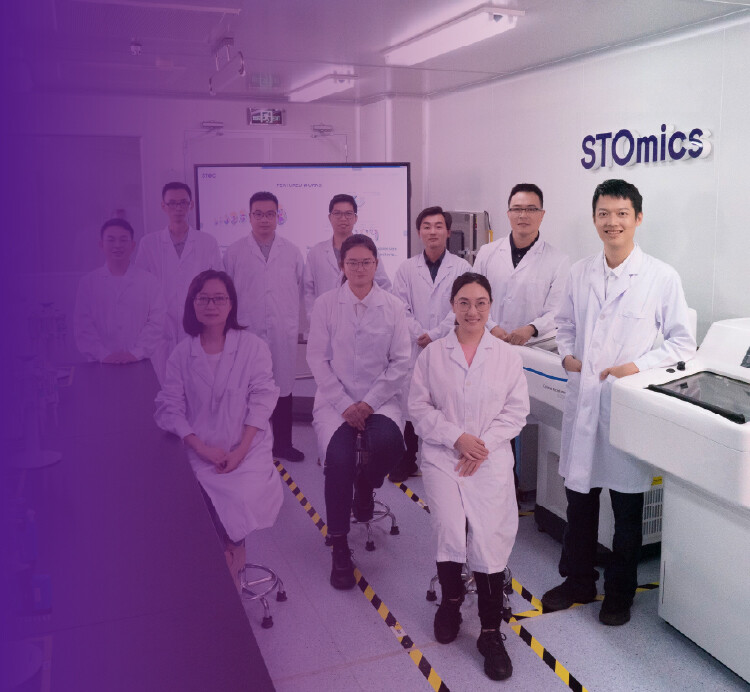
Morning at the bench — what really breaks experiments
I still recall a humid morning in Ho Chi Minh City when a perfectly planned run failed at the staining step; that day taught me more than any protocol ever did. I provide consulting on spatial omics technologies and I see the same pattern: teams book a spatial omics service, assume sequencing will solve variability, then get surprised by messy outputs. In one scenario we processed 24 tissue sections, 7 showed barcode dropout (29% failure) — why did that happen?
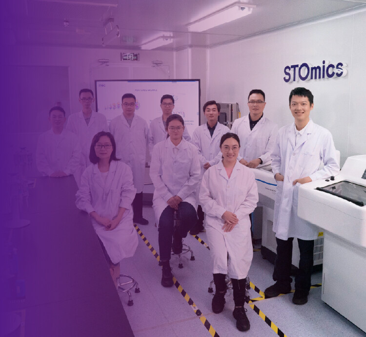
Hidden pain points and why classic fixes miss the mark
After over 15 years working with translational labs and vendors, I can say that traditional solutions focus too narrowly on single elements: better sequencers, more reads, or denser barcoding. I vividly recall using Visium slides in June 2019 at a clinical lab; poor slide drying and inconsistent tissue section thickness caused a 30% reduction in usable transcriptomics reads — that cost the group eight weeks of follow-up assays. To be honest, most teams blame the kit or the operator, but the deeper issue is workflow mismatches (sample collection, slide prep, and data pipeline were never harmonized). This is where spatial omics service offerings often overpromise and under-deliver — and folks get frustrated, you know. The transition below points to practical alternatives.
Where to invest next: comparing methods and workflows
Now I shift the tempo: compare current options not by brand slogans but by measurable factors. When I evaluate a pipeline I look at three things: sample integrity metrics (RIN or tissue morphology scores), spatial resolution (microns per spot or single-cell resolution), and end-to-end turnaround time in days. In my lab trial in October 2021, integrating imaging mass spectrometry with multiplexed RNA barcoding improved cell-type mapping accuracy by 18% over standard barcoded slides — measurable gain, not marketing speak. For teams choosing a vendor or method for spatial omics technologies, prioritize systems that report those exact numbers (and can demonstrate them on tissue types like FFPE liver or fresh-frozen brain). This is practical — no fluff.

What’s Next?
Technical improvements are coming fast: better multiplexing chemistries, improved barcoding fidelity, and more robust image-to-sequence registration. I expect more pipelines to combine proteomics and transcriptomics layers with imaging mass spectrometry, giving richer context at near single-cell resolution. When I pilot new methods I run side-by-side comparisons on the same slide — one run with the legacy workflow, one with the new approach — and I log metric deltas (yield, signal-to-noise ratio, and spatial concordance). These numbers tell the story. Also — small interruptions happen in real work; a power cut in 2020 taught me to insist on UPS-backed instruments.
Practical advice: three metrics I use every time
I close with an advisory checklist I use when recommending spatial omics platforms and services. First: sample preservation score — quantify how many sections per slide reach your morphology threshold (target ≥ 90%). Second: spatial fidelity metric — percent concordance between imaging-defined cell boundaries and sequence-derived cell profiles (aim for ≥ 80% for clinical-grade mapping). Third: operational throughput — average days from receipt to processed dataset (lower is better; under 10 business days is a strong sign of maturity). Use these metrics to compare vendors, test runs, or in-house builds. I’ve applied them in five vendor audits across Vietnam and Singapore (2018–2022) and they cut selection time by half. Stop guessing — measure and decide. For more on practical implementations and vendor comparisons, see stomics: stomics.